


To date, science is known for 280 types of worms that can develop and live in the human body, parasitized in various organs and tissues.The frequency of human infection depends on the climatic and socio-economic conditions of the specific areas (in the subjects in the underdeveloped countries, especially in the tropical and subtropical zones, the level of parasitic infections is much higher than in economically developed conditions).
Methods of Human Infection with Helmints:
- Biogelmintosis (infection of animals).
- Fertőoin Helmintoses (transmitted from person to person).
- Geogelmintoses (diseases caused by one of the vital cycles on the earth).
Factors that influence the manifestations of helminthiases
Method of penetrating a parasite into the body:
- The degree of adaptation of helmin to the human body;
- The density (quantity) of the parasitic individuals;
- The habitat of the worm (tissue parasites live in the thickness of the soft tissues and the cleaning of the hollow organs).Some helmin has forms of educational and tissue in different phases.The larva and developing stages of worms usually cause more pronounced abnormal changes.
In the absence of infections, the number of adult parasites in the human body does not increase.This property significantly distinguishes the halminth invasions from diseases caused by bacteria, viruses, fungi and simple organisms.
Worms in people: symptoms
Helminthosis is a disease characterized by 2 stages of the course (acute, two weeks to two months) and chronic (several months for several years).
Symptoms of acute phase of helminthiasis
The first signs of the disease may occur at different times (most often after 2-3 weeks, with 2-3 days of ascaridosis, and the incubation period for phaylairiosis may last 6-18 months).
The most typical symptom in the acute stage of parasitic invasion is an allergic reaction (antibodies are generated by the antigens of the migrant larvae of parasites).Itchy rashes prone to returning paths often increase on the returning track.In addition, the wandering larvae of the parasite can cause pain in the chest, cough, drowning, stool disorders, nausea and vomiting.
At the same time, acute phase of helminthiasis is accompanied by more severe disorders (severe forms of pneumonia, hepatitis, allergic myocarditis, hepatosterogalia (liver and spleen growth).
In the blood, the amount of eosinophils (eosinophilia) increases and interferes with the normal quantitative ratio between protein motions (dysproteinemia).
Signs of chronic helminthiasis
The symptoms of the chronic phase depend directly on which organ "populated" parasites and their size and quantity play an important role.
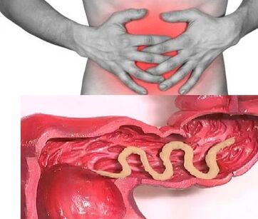
So, if the unique individuals are a parasite in the bowel, the disease can be asymptomatic (in the case of infection with very large parasites).The characteristic properties of the chronic phase of the intestine are the dyspeptic disorders.In children, the acenoarotic and pain syndrome is better pronounced.Massive invasion ascarides, bowel obstruction, mechanical jaundice and pancreatitis.
With the consumption of all substances needed for their vital activity from the body of the host, the helmink causes indigestion disorders, violations of the absorption of vitamins, minerals, carbohydrates, proteins and fats.At the same time, the products of worms inhibit normal intestinal microflora and reduce body immune power.
In people with helminthias, due to the increased process of weakened immunity and the increased process of cell distribution (consequences of constant restoration of tissue parasites), the risk of malignant tumors increases significantly.
The parasite types of helmints in the human body
Human Helminthiasis pathogens are 2 types of worms: round (nematodes) and flat (ribbon and saucers).
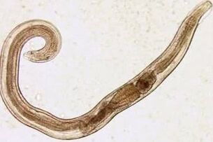
Round worms
Luggage
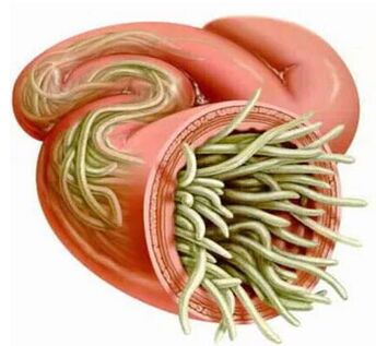
The parasites of the causes of enterobiosis are small (up to 10 mm) thin cavities with greyish-white painting.The infection occurs in a nutritious way (through the mouth).The reason for this is the dirty hands.The eggs of the parasite may be on the ground, the wool of infected animals, the unwashed vegetables and fruits, etc.At the same time, in the case of enterobiosis, the case of self -extension (especially in children) that occur as a result of the combing of the areas and the subsequent eggs.The cutting larva develops within two weeks in the digestive tract.When becoming an adult, the worm parasitizes in the lower and upper parts of the colon.
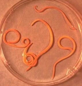
Ascarida
Ascaride is a large parasite for red-yellow spindle-shaped, 40 cm (female) and 15-25 cm (men) adult.Without a suction cup or other recording device, the ascaride can move independently towards food masses.Eggs laid down by the female parasite are distinguished by stool.
Acidic infection is the swallow of mature eggs, water or unwashed vegetables and fruits with soil particles.After the eggs penetrate the gut, the mature larvae came out of them.The introduction to the intestinal wall is then reached by blood flow and falls into the lungs.Through the lungs alveolia, the larva of the ascarida penetrates the oral cavity again through the respiratory system.After repeated swallowing, the parasite reaches the small intestine where it becomes adult.The worm lives for 12 months, then dies and stands out with the stool.In the bowel of one owner, one can live both or hundreds of people.
Vlashev
Vlasov, a trichocephalosis causal agent, a white helmin that parasitizes in the initial part of the colon and reaches 4-5 cm.The parasite is nourished by the blood and tissues of the rectal mucosa.
On the walls of the intestine, the eggs that are sealed by the female by the female come out with the stool.They are developed in the environment (optimally in the soil).Eggs with parasitic larvae are penetrated into the body with dirty hands, water or unwashed vegetables and fruits.
Trichinella
The pathogen of trichinellosis is a small round Helmin, which reaches 2-5 mm.The infection occurs when the use of poorly roasted meat (pork, bear puppies, wild boar).Penetrating the intestine, the parasitic larva is matured in 3-4 days to the state of the sexually mature individual.The life expectancy of the worm is 40 days and then the parasite dies.Under the guidance of the intestinal wall, the larvae penetrate into the bloodstream and carry it in every organ of the human body, settled in the muscles.In this case, the respiratory and facial muscles and the muscles of the limbs are most often affected.
In the early days after invasion, patients complain of abdominal pain.Then approx.After 2 weeks, body temperature rises to 39-40 s, itchy rashes occur on the skin, the muscle pain develops and the face swells.During this period, in the event of enormous infection, it is a significant risk of mortality.After about a month, healing occurs.The parasite is capsulated in a spiral form and dies within two years.
Ankylostoma and no -Core
These two parasites are similar to biological characteristics and diseases caused.This is customary to combine them as a general name (ankylostoma).Worms of 10-15 mm are parasitized in 12-P.intestines.It should be noted that this is one of the most common but at the same time rarely identified parasites.Worms of worms penetrate into the human body through the skin when they come into contact with infected soil.In addition, entry into the bloodstream, like the Ascarides, migrates into the lungs and then through the bronchi, through the jumping sputum into the digestive system.The anquilostoma is parasitized in the intestine, attached to the intestinal wall.The parasite that eats exclusively with blood bites is pierced by the blood vessels and injected an anti -sensed component there.During the day, an adult is able to absorb an average of 0.05-0.35 ml of blood.Therefore, the most typical symptom of this halminthiasis is the change in iron deficiency and the proportion of protein refractions (dysproteinemia).
Flat worms
Wide strip
This is one of the biggest helmin that reaches 10-20 meters.The disease caused by this parasite is called dipillobotriosis.The worm development cycle begins with freshwater fish or crustaceans.The larva enters the human body, which is the ultimate owner of a wide ribbon with caviar or infected fish fillets.By reaching the small intestine, the parasite is attached to the wall and increases for 20-25 days to a sexually mature individual.
Liver
The parasite that causes opisthorchiasis is a flat worm that reaches 7-20 mm length.It should be noted that more than 50% of the infection of the liver saucer (also known as cat bikonomer) falls on the inhabitants of Russia.The parasitic larvae begin to form after the eggs fall into fresh water (swallowed from the snails).They then penetrate into the fish body (carp, intersection, bream, kick).Human infection occurs when eating an infected fish that has not undergone enough heat treatment.The larvae from the small intestine penetrates into the bile ducts and the gallbladder and secured with two sucks.
Bull and pork
These parasites are almost the same 5-6 meters long.Infection of tusiarinhosis and tussis is due to the use of Finnish -infected cattle or pork (one of the intermediate forms of helminthiasis).The visible Finnish, which is shown in the form of whitish bubbles, reaching the size of 0.5 cm, is connected to the wall of a person and develops as an adult within 3 months.The striptease parasite, which consists of more than 2000 segments, is constantly increasing.At the same time, the final segments containing eggs descend and independently move along the colon to the anal opening, then leak from the anal or stand out into the outer environment beside the stool.The most typical symptoms of helminthiasis are the digestive system.
Echinococcus
For this parasite, man is an intermediate host.The worm is parasitized in the human body in its Finnish form.The last owner of echinococcus is a wolf, dog or cat.The infection occurs in a nutritious way in contact with animals and environmental objects, with a handful of echinococcal eggs.After entering the intestine, they develop oncospheres (six -black larvae).They penetrate the bloodstream from the intestine and carry it through the whole body.
Alley
This parasite, which is considered to be a variety of echinococcus, is one of the most dangerous causes of helmintia (alveococcosis), which is similar in the severity of cirrhosis and liver cancer.The infection is penetrated into the intestines by penetrating the oncosphere (egg mature larvae).There, the embryo comes out of the egg and introduced into the intestinal walls into the bloodstream.In addition, blood flow spreads the parasite on all tissues and organs of the body (most often localized in the liver).It is there that the main phase of development begins on the larvae (a multi -camera bubble, laurelocyst).Each ventricle contains a parasite embryo head that continues to develop gradually.Lavrocalists are very aggressive formations that are constantly growing due to growing bubbles and can germinate in the liver, such as cancer metastases.
Diagnosis of helminthiasis
Diagnosis of Helmintes invasions includes the following events:
- Thorough collection of anamitesis to help you learn the potential causes of the infection;
- Stools, blood, 12p intestines, rectum and perianal mucosa, muscle tissue, lung sputum, bile laboratory examination.During the analysis, eggs, segments or fragments of parasites can be detected.At the same time, the increased content of eosinophils in the blood is also a sign of the presence of helminthiasis.
- Serological examinations (ELISU, RSC, indirect agglutination, immunofluorescence analysis, etc.) in the diagnosis of diseases caused by larva stages or tissue parasites.
- Ultrasound, CT and endoscopic tests are required to identify the liver tissue.
Worms in people: treatment
During the acute phase of parasitic infection, the patient is detoxified and desensitizing therapy is prescribed.During the severe course of the disease (liver trematodosis, trichinellosis), glucocorticoids are used according to medical indications.
Specific anthelmintic chemotherapeutic agents are prescribed as specific therapeutic drugs, taking into account the nature of the pathogen.
In parallel, the patient is recommended to take antihistamines and enterosorbens.The last stage of treatment includes the use of normalizing probiotics for the intestinal microflora.
A special economical diet is also prescribed (foods should be easily digestible and have little fat).
During anthelmintic therapy, the patient should strictly adhere to personal hygiene (to avoid re -infection).At the same time, with many helminthiasis, all family members and persons infected must be treated.